Ovary/Oocyte
Poster Session B
(P-272) In Vitro Differentiation of Bovine Embryonic Stem Cells Toward Progenitors of Bipotential Gonad-like Somatic Cells
Thursday, July 18, 2024
8:00 AM - 9:45 AM IST
Room: The Forum

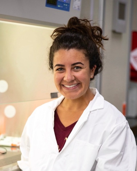
Juliana I. Candelaria, MS
Graduate Student
University of California Davis
DAVIS, California, United States
Poster Presenter(s)
Abstract Authors: Juliana I. Candelaria, Carly Guiltinan, Ramon C. Botigelli, Anna C. Denicol
Abstract Text: Somatic cells of the mammalian gonad originate from the mesoderm, which differentiates into the intermediate mesoderm (IM), coelomic epithelium (CE), and then bipotential gonad (BG) via cell signaling by specific morphogens. Research in human gonadal cell specification remains hampered by difficulty accessing embryos; therefore, stem cell-based models have been used to recapitulate stepwise gonadal cell differentiation in vitro. However, reports of successful cell induction into BG-like cells are still limited. Hence, in vitro differentiation of bovine embryonic stem cells (bESCs) into BG-like cells presents opportunities to understand mechanisms of early gonad development in a large mammal model. We hypothesized that bESCs would differentiate into IM and early CE (eCE) after sustained WNT activation, short duration of activin A (Nodal) and bFGF (FGF) signaling, and low concentrations of BMP4 during in vitro culture. In experiment 1 (n = 3 replicates), bESCs (p19-26) were cultured for 48 h in mesoderm induction medium (MIM) containing 3 µM CHIR99021 (GSK3 inhibitor/WNT activator) and 70 ng/mL activin A. Cells were then further cultured without or with 10 ng/mL bFGF starting at 48, 72, or 96 h and harvested at 120 h. In experiment 2 (n = 3 replicates), we sought to determine if low bFGF exposure with BMP4 synergistically upregulated IM and eCE markers. As in experiment 1, bESCs were cultured in MIM for 48 h and then supplemented with 0, 1, 10, or 20 ng/mL BMP4 for an additional 72 h with a pulse of 10 ng/mL bFGF during the final 24 h of BMP4 exposure. Cells were harvested at 120 h and subjected to RT-qPCR to examine expression of IM and eCE/bipotential gonad transcripts. After 48 h of mesoderm induction, LHX1 (early IM marker) showed peak expression in both experiments (P < 0.001). After 72 h of additional culture, LHX1 became downregulated independently of bFGF or BMP4 supplementation (P < 0.0001), aligning with in vivo temporal expression patterns. Likewise, 72 h after mesoderm induction, OSR1 (IM marker) and WT1 (eCE marker) were upregulated similarly across bFGF and BMP4 regimens. We found no difference in GATA4 (CE/BG marker) expression across bFGF regimens, however 10 and 20 ng/mL of BMP4 increased GATA4 compared to bESCs (P < 0.05). PAX3 (paraxial mesoderm marker) expression was low and similar to bESCs in all culture conditions in both experiments, indicating appropriate suppression of the paraxial mesoderm linage. We also found that at the end of the 120 h culture period, FOXF1 [lateral plate mesoderm (LPM) marker] was upregulated in the absence or after short duration exposure to bFGF (24 or 48 hours of culture; P < 0.05). There was a dose-dependent increase in FOXF1 (P < 0.05) in response to BMP4, indicating a shift towards the LPM in response to an increased gradient of BMP4 as it occurs in vivo. We corroborated our findings of WT1 expression by demonstrating WT1 protein expression in bESC following exposure to BMP4 with bFGF via immunocytochemistry. Together, these data suggest that 1) bESCs are being driven towards the IM and eCE state by 120 h of culture with constant WNT activation, hence show potential to be further driven towards a somatic BG-like state; 2) high BMP4 with short bFGF exposure may induce the LPM; and 3) paraxial mesoderm fate is not induced by these culture conditions. Financial support: NIH F31 Pre-doctoral fellowship.
Abstract Text: Somatic cells of the mammalian gonad originate from the mesoderm, which differentiates into the intermediate mesoderm (IM), coelomic epithelium (CE), and then bipotential gonad (BG) via cell signaling by specific morphogens. Research in human gonadal cell specification remains hampered by difficulty accessing embryos; therefore, stem cell-based models have been used to recapitulate stepwise gonadal cell differentiation in vitro. However, reports of successful cell induction into BG-like cells are still limited. Hence, in vitro differentiation of bovine embryonic stem cells (bESCs) into BG-like cells presents opportunities to understand mechanisms of early gonad development in a large mammal model. We hypothesized that bESCs would differentiate into IM and early CE (eCE) after sustained WNT activation, short duration of activin A (Nodal) and bFGF (FGF) signaling, and low concentrations of BMP4 during in vitro culture. In experiment 1 (n = 3 replicates), bESCs (p19-26) were cultured for 48 h in mesoderm induction medium (MIM) containing 3 µM CHIR99021 (GSK3 inhibitor/WNT activator) and 70 ng/mL activin A. Cells were then further cultured without or with 10 ng/mL bFGF starting at 48, 72, or 96 h and harvested at 120 h. In experiment 2 (n = 3 replicates), we sought to determine if low bFGF exposure with BMP4 synergistically upregulated IM and eCE markers. As in experiment 1, bESCs were cultured in MIM for 48 h and then supplemented with 0, 1, 10, or 20 ng/mL BMP4 for an additional 72 h with a pulse of 10 ng/mL bFGF during the final 24 h of BMP4 exposure. Cells were harvested at 120 h and subjected to RT-qPCR to examine expression of IM and eCE/bipotential gonad transcripts. After 48 h of mesoderm induction, LHX1 (early IM marker) showed peak expression in both experiments (P < 0.001). After 72 h of additional culture, LHX1 became downregulated independently of bFGF or BMP4 supplementation (P < 0.0001), aligning with in vivo temporal expression patterns. Likewise, 72 h after mesoderm induction, OSR1 (IM marker) and WT1 (eCE marker) were upregulated similarly across bFGF and BMP4 regimens. We found no difference in GATA4 (CE/BG marker) expression across bFGF regimens, however 10 and 20 ng/mL of BMP4 increased GATA4 compared to bESCs (P < 0.05). PAX3 (paraxial mesoderm marker) expression was low and similar to bESCs in all culture conditions in both experiments, indicating appropriate suppression of the paraxial mesoderm linage. We also found that at the end of the 120 h culture period, FOXF1 [lateral plate mesoderm (LPM) marker] was upregulated in the absence or after short duration exposure to bFGF (24 or 48 hours of culture; P < 0.05). There was a dose-dependent increase in FOXF1 (P < 0.05) in response to BMP4, indicating a shift towards the LPM in response to an increased gradient of BMP4 as it occurs in vivo. We corroborated our findings of WT1 expression by demonstrating WT1 protein expression in bESC following exposure to BMP4 with bFGF via immunocytochemistry. Together, these data suggest that 1) bESCs are being driven towards the IM and eCE state by 120 h of culture with constant WNT activation, hence show potential to be further driven towards a somatic BG-like state; 2) high BMP4 with short bFGF exposure may induce the LPM; and 3) paraxial mesoderm fate is not induced by these culture conditions. Financial support: NIH F31 Pre-doctoral fellowship.
